Digital Dental X-Rays
What Are Digital Dental X-Rays?
Dental X-rays have been an essential tool in dentistry for decades, aiding dentists in detecting various oral health issues that may not be visible during a routine dental examination. By utilizing low levels of radiation to penetrate oral tissues and create detailed images of teeth, gums, and bones, X-rays provide crucial insights into a patient’s oral health, assisting in the early identification and treatment of dental problems.
Digital dental X-rays have emerged as a modern alternative to the conventional film-based method. In this technology, a digital sensor is used to capture the X-ray images, which are then processed and displayed on a computer screen. The transition to digital X-rays has significantly enhanced the diagnostic process, benefiting both dental practitioners and patients alike. Before you deciding on whether Digital Dental X-Rays are right for you, there are some things you should know:
- Who Needs Digital Dental X-Rays?
- What Are The Different Types Of Digital Dental X-Rays?
- What Are The Advantages Of Digital Dental X-Rays?
- How Much Does Digital Dental X-Rays Cost?
- What Are The Steps In The Digital Dental X-Rays Procedure?
- Can I Have Digital Dental X-Rays If I Am Pregnant?
If you have any further questions about Digital Dental X-Rays or other dental services offered at Atlas Dental, please contact us.

Free phone consultation
Have questions about CBCT Scans? Schedule a free phone consultation with our Toronto dentist.

5 star google reviews
Our patients love us! See for yourself why more and more people are choosing Atlas Dental for their CBCT Scans.

Book CBCT Scan Online
We provide quick and convenient CBCT Scan Service in Toronto. Book Online.
Who Needs Digital Dental X-Rays?
Digital dental X-rays are an integral part of routine dental care and are recommended for a wide range of patients. The decision to utilize X-rays depends on various factors, including a person’s oral health history, age, risk of dental issues, and specific symptoms they may be experiencing. Let’s explore the different categories of patients who may benefit from digital dental X-rays:
- New Patients: For individuals visiting a dental office for the first time, digital dental X-rays are often part of the initial examination process. These X-rays provide a comprehensive view of the patient’s oral health, allowing the dentist to identify any existing dental problems, assess the condition of the teeth and supporting structures, and establish a baseline for future comparisons.
- Routine Check-ups: Regular dental check-ups typically involve periodic X-rays to monitor the overall oral health and identify any potential concerns early on. The frequency of X-rays varies depending on the patient’s age, oral health, and risk of developing dental issues. In most cases, adults may require X-rays every 1 to 2 years, while children or individuals with higher risk factors may need them more frequently.
- Dental Concerns: When patients present specific dental symptoms or complaints, digital dental X-rays play a crucial role in diagnosing the underlying issues. Whether it’s unexplained tooth pain, changes in the gum tissue, or suspected dental decay, X-rays provide valuable information that assists the dentist in devising an appropriate treatment plan.
- Orthodontic Treatment: Orthodontists use digital dental X-rays to assess the alignment and positioning of teeth, jawbone structure, and other factors necessary for developing an orthodontic treatment plan. These X-rays aid in determining the most suitable orthodontic appliances, such as braces or Invisalign clear aligners, and tracking progress throughout the treatment process.
- Preparatory Procedures: Before certain dental procedures, such as dental implants or extractions, digital dental X-rays are essential to assess the underlying bone structure and ensure a successful and safe treatment outcome.
- Periodontal (Gum) Disease Evaluation: X-rays help dentists evaluate the extent of gum disease and bone loss in patients with periodontal issues. This information is crucial for determining the appropriate periodontal treatment needed to address the condition effectively.
It’s important to note that while digital dental X-rays are widely used, dentists follow strict guidelines and principles of ALARA (As Low As Reasonably Achievable) when it comes to radiographic imaging. This means that X-rays are only prescribed when the benefits of obtaining the diagnostic information outweigh the potential risks associated with radiation exposure. If you have further questions about Digital Dental X-Rays, please contact us.
What Are The Different Types Of Digital Dental X-Rays?
There are several different types of digital dental X-rays, each serving specific purposes and providing valuable insights into different aspects of a patient’s oral health. Here are the most common types of digital dental X-rays:
- Intraoral X-Rays: Intraoral X-rays are the most frequently used type of dental X-rays. These images are taken with the X-ray sensor placed inside the mouth, providing a close-up view of individual teeth and their surrounding structures. Intraoral X-rays can be further classified into the following subtypes:
- Bitewing X-Rays: These X-rays capture the upper and lower teeth in a single view when the patient bites down on a specialized film or sensor holder. Bitewing X-rays are commonly used to detect dental caries (cavities) between teeth and assess the fit of dental restorations.
- Periapical X-Rays: Periapical X-rays focus on a specific tooth, showing its entire length from the crown to the root and the surrounding bone. They are useful for diagnosing issues related to the tooth’s root, such as infections or abscesses.
- Extraoral X-Rays: Unlike intraoral X-rays, extraoral X-rays are taken from outside the mouth and capture a broader view of the head and jaw. They are especially helpful in assessing the overall jaw structure and positioning of the teeth. The most common types of extraoral X-rays include:
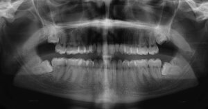
- Panoramic X-Rays: Panoramic X-rays provide a sweeping view of the entire mouth, including all the teeth, upper and lower jaws, temporomandibular joints (TMJ), and surrounding structures. They are useful for evaluating impacted teeth, jaw problems, and planning orthodontic or surgical treatments.
- Cephalometric X-Rays: Cephalometric X-rays focus on the side profile of the head, capturing the bones of the face and skull. Orthodontists often use these X-rays to plan orthodontic treatments and assess skeletal growth patterns.
- Cone Beam Computed Tomography (CBCT): CBCT is an advanced digital imaging technique that provides three-dimensional images of the oral structures. It uses a cone-shaped X-ray beam to capture detailed images of teeth, bone, nerves, and soft tissues. CBCT is particularly valuable in dental implant planning, oral surgery, endodontics (root canal treatment), and complex orthodontic cases.
Each type of digital dental X-ray serves a specific diagnostic purpose and complements the others in providing a comprehensive assessment of a patient’s oral health. The choice of which X-rays to use depends on the patient’s individual needs, symptoms, and the dental professional’s diagnostic requirements. Digital X-rays have transformed the field of dentistry, enabling dental practitioners to diagnose dental issues more accurately and tailor treatment plans to meet each patient’s unique needs. If you have further questions about Digital Dental X-Rays, please contact us.
What Are The Advantages Of Digital Dental X-Rays?
Digital dental X-rays offer a myriad of benefits over their conventional film-based counterparts, making them an indispensable tool in modern dentistry. Let’s delve into the key advantages that have propelled the widespread adoption of digital dental X-rays:
- Reduced Radiation Exposure: One of the most significant advantages of digital dental X-rays is the significant reduction in radiation exposure compared to traditional X-rays. Digital X-ray sensors are more sensitive and require much less radiation to produce high-quality images. This makes the process safer for patients, and it aligns with the ALARA principle, ensuring that the X-rays are kept “As Low As Reasonably Achievable.”
- Immediate Image Acquisition: Unlike traditional X-rays that involve manual film processing, digital dental X-rays provide instant image acquisition. The X-ray images are captured by the sensor and displayed on a computer screen within seconds. This real-time visualization enables dentists to assess and discuss the results with patients immediately, leading to quicker diagnosis and treatment planning.
- Enhanced Image Quality: Digital dental X-rays offer superior image clarity and resolution compared to traditional film X-rays. The high-quality images provide dentists with better visibility of dental structures, allowing them to detect dental issues with greater precision. This improved accuracy leads to better diagnosis and more effective treatment outcomes.
- Image Manipulation and Enhancement: Digital X-ray images can be easily manipulated and enhanced using specialized software. Dentists can zoom in, adjust brightness and contrast, and even annotate the images to highlight specific areas of concern. This versatility aids in better communication with patients, as dentists can explain their findings in a more accessible manner.
- Eco-Friendly and Cost-Effective: Digital dental X-rays eliminate the need for chemical processing and film storage, making them environmentally friendly and reducing overall operational costs for dental practices. Additionally, since digital X-ray images are stored electronically, the need for physical storage space is minimized, streamlining record-keeping and improving office efficiency.
- Easy Image Sharing and Transmission: Digital X-ray images can be easily shared and transmitted electronically between dental professionals, specialists, or for consultation purposes. This seamless sharing of information facilitates collaborative dental care and ensures that all members of the dental team are well-informed about a patient’s oral health status.
- Patient Education and Involvement: With the ability to display X-ray images on a computer screen, dentists can engage patients more actively in their treatment process. Visual aids enable patients to better understand their oral health conditions, treatment options, and the importance of preventive care, fostering a sense of empowerment in their own dental care journey.
- Reduced Appointment Time: The immediate availability of digital X-ray images reduces the need for additional appointments solely for X-ray evaluation. Dentists can review the images during the same visit, expediting the diagnosis and treatment planning process.
Digital dental X-rays have become an indispensable tool in modern dentistry, offering advantages such as reduced radiation exposure, real-time imaging, enhanced image quality, and eco-friendliness. With improved diagnostic capabilities and patient engagement, digital X-rays play a vital role in ensuring comprehensive and efficient dental care. If you have further questions about Digital Dental X-Rays, please contact us.
Cost of Dental X-Rays Images
The cost of Dental X-Ray Images will depend on the type of image taken as well as the number of images. The codes relevant to dental x-rays in the Ontario Dental Association’s Suggested Fee Guide appear as follows:
Radiographs, Periapical
- 02111 – Single image: $40
- 02112 – Two images: $47
- 02113 – Three images: $54
- 02114 – Four images: $61
- 02115 – Five images: $72
- 02116 – Six images: $80
- 02117 – Seven images: $90
Radiographs, Bitewing
- 02141 – Single image: $42
- 02142 – Two images: $52
- 02143 – Three images: $63
- 02144 – Four images: $73
Radiographs, Regional/Localized
- 02101 – Radiographs, Complete Series (minimum of 12 images incl. bitewings): $162
- 02102 – Radiographs, Complete Series (minimum 16 images, incl. bitewings): $175
Radiographs, Panoramic
- 02601 – Single images: $82
Dental X-Rays are considered a basic service under all dental insurance plans and should be covered to your maximum insurable limit, but be sure to find out from your dental insurance plan provider how much you are eligible for before going ahead with dental treatment. Our fees are consistent with the ODA Fee Guide.
For patients without dental insurance, Atlas Dental is pleased to offer dental financing through Dentalcard. Affordable payment plans start at 7.95% for terms of 6 months to 6 years. To learn more about Dentalcard dental treatment financing, follow this link.
What Are The Steps In The Digital Dental X-Rays Procedure?
The digital dental X-rays procedure is a straightforward and efficient process that allows dentists to capture detailed images of a patient’s oral structures. The method involves several simple steps, ensuring minimal discomfort and radiation exposure for the patient. Let’s explore the typical steps involved in the digital dental X-rays procedure:
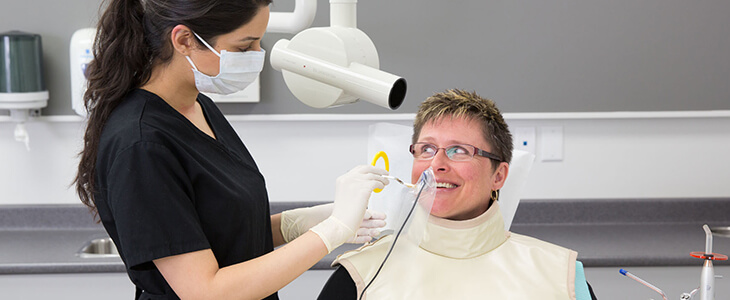
- Preparation and Safety Measures: Before the X-ray examination, the dental professional will take necessary precautions to ensure the patient’s safety and comfort. This may involve providing a lead apron and thyroid collar to shield sensitive areas of the body from radiation exposure. The patient will be positioned appropriately to ensure the accurate capture of X-ray images. The dental assistant or hygienist may help position the X-ray sensor in the mouth to get the desired views.
- X-Ray Image Capture: The dental professional will use a small, digital X-ray sensor, which resembles a small, flat device, to capture the images. This sensor is placed inside the mouth and positioned strategically to capture different angles and views of the teeth and surrounding structures. The dentist will then leave the room and activate the X-ray machine, which emits a brief burst of radiation to capture the images. The radiation exposure is minimal, thanks to the sensitivity of digital sensors.
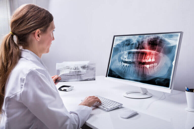
- Image Processing: Once the X-ray images are captured, they are processed immediately and appear on the computer screen in real-time. The quick processing allows the dentist to review the images promptly, eliminating the wait time associated with film-based X-ray processing.
- Image Review and Analysis: The dentist will carefully examine the X-ray images on the computer screen. They will look for any signs of dental decay, gum disease, infections, abnormalities, or other oral health issues. The high-quality digital images provide better visibility of dental structures, allowing the dentist to make a more accurate diagnosis.
- Patient Consultation: After reviewing the X-ray images, the dentist will discuss their findings with the patient. They may use the images to explain the diagnosed conditions, treatment options, and the importance of maintaining good oral hygiene. Patient education plays a vital role in ensuring that the patient is actively involved in their dental care decisions.
- Image Storage and Record-Keeping: Digital dental X-ray images are stored electronically in the patient’s dental records. This electronic storage eliminates the need for physical film storage and provides easy access to the images for future reference and comparison.
- Follow-Up and Treatment Planning: Based on the X-ray findings, the dentist will develop a personalized treatment plan, if necessary, to address any detected dental issues. This plan may include further dental treatments, preventive measures, or recommendations for maintaining optimal oral health.
The digital dental X-ray procedure ensures an efficient and accurate diagnostic process for dental professionals while prioritizing patient safety and convenience. If you have further questions about Digital Dental X-Rays, please contact us.
Can I Have Digital Dental X-Rays If I Am Pregnant?
Pregnancy is a time when women become particularly concerned about the potential risks of any medical procedures, including dental X-rays. The question of whether a pregnant woman can have digital dental X-rays depends on several factors, including the stage of pregnancy, the necessity of the X-rays, and the specific dental concern being addressed:
- Necessary Dental Treatment: If you are pregnant and require dental treatment, it is essential to inform your dentist about your pregnancy. Some dental issues may require X-rays to determine the best course of action. However, non-urgent X-rays are typically postponed until after the pregnancy to avoid unnecessary exposure to radiation.
- Lead Apron and Thyroid Collar: If a dental X-ray is deemed necessary during pregnancy, your dentist will take extra precautions to protect both you and your baby. You will be provided with a lead apron to wear during the X-ray procedure. The lead apron acts as a shield, protecting the abdomen and pelvic area from radiation exposure. Additionally, a thyroid collar may be used to protect the thyroid gland, which is situated in the neck area and is also sensitive to radiation.
- Limited Exposure and ALARA Principle: Dental professionals adhere to the ALARA principle (As Low As Reasonably Achievable) when taking X-rays on pregnant patients. This means that the X-ray exposure is kept to the minimum necessary to obtain diagnostic information. Digital dental X-rays already require lower radiation levels compared to traditional film X-rays, contributing to the overall reduction in radiation exposure.
- Discussing Risks and Benefits: If there is any doubt or concern about the necessity of dental X-rays during pregnancy, it is essential to have an open and honest conversation with both your dentist and obstetrician. Together, they can assess the risks versus the benefits of the X-rays and make an informed decision regarding your dental care.
- Timing of X-Rays: In general, dental X-rays are typically avoided during the first trimester of pregnancy, as this is a critical time in fetal development. If possible, any necessary X-rays are postponed until the second or third trimester when the fetus is more developed and less susceptible to potential risks.
- Alternatives and Safe Practices: Whenever possible, alternative diagnostic methods or temporary treatment options may be considered to manage dental issues during pregnancy. Good oral hygiene practices, regular dental check-ups, and preventive care play a vital role in maintaining oral health during pregnancy.
If you are pregnant or planning to become pregnant, be sure to inform your dental professional so that they can provide you with the best and safest care tailored to your specific needs. If you have further questions about Digital Dental X-Rays, please contact us.
We also think you’ll like…

Bicon Dental Implants
Bicon Dental Implants What Are Bicon Dental Implants? Bicon is a well-respected dental implant company known for its innovative approach to implantology and its unique
Wisdom Tooth Removal Consent Form
Wisdom Tooth Removal Consent Form There will be some pain, swelling and bleeding following a tooth extraction. This may require pain-relieving medication. Bleeding is usually
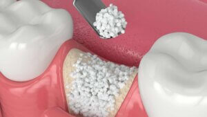
Alveolar Bone Preservation
Alveolar Bone Preservation What Is Alveolar Bone Preservation? Alveolar bone preservation is a dental procedure that involves placing a bone graft material into the socket

Biohorizons Dental Implants
Biohorizons Dental Implants What Are BioHorizons Dental Implants? BioHorizons is a prominent dental implant company known for its expertise in the design, manufacture, and distribution
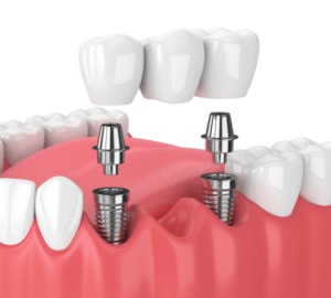
Dental Implant Bridge Home Care Instructions
Dental Implant Bridge Home Care Instructions What You Should Do After Receiving Your Dental Implant Bridge A Dental Implant Bridge is a prosthetic device used

Dental Pain Medication
Dental Pain Medication What Is Dental Pain Medication? Dental pain can range from a mild discomfort to intense throbbing, making even the simplest tasks unbearable.